

The Thoracic Duct: An Osteopathic Exploration of Anatomy, Respiration and Lymphatic Function
Oct 02, 2026Most osteopaths will have studied the thoracic duct during their anatomical education. We know that it is the largest lymphatic vessel in the body, that it returns lymph to the venous circulation and that it travels from the abdomen through the thorax before terminating somewhere around the root of the neck.
And for many of us, that is probably where our knowledge of the thoracic duct ends.
But when we start looking at this structure in greater detail, we discover something far more interesting than the simple green line illustrated in our anatomy textbooks.
The thoracic duct is a living, contractile and remarkably variable vessel. It travels through regions that are constantly changing with respiration, movement and pressure. Its development helps explain its unusual course and its relationships with the diaphragm raise some fascinating questions about lymphatic physiology.
For me, it is a wonderful example of why anatomy becomes so much more useful when we stop studying structures in isolation and start exploring the relationships between them.
Where does the thoracic duct begin?
The traditional anatomical description places the beginning of the thoracic duct at the cisterna chyli.
This is usually illustrated as an elongated lymphatic sac lying anterior to the upper lumbar vertebrae, commonly around L1 or L2, close to the right side of the abdominal aorta.
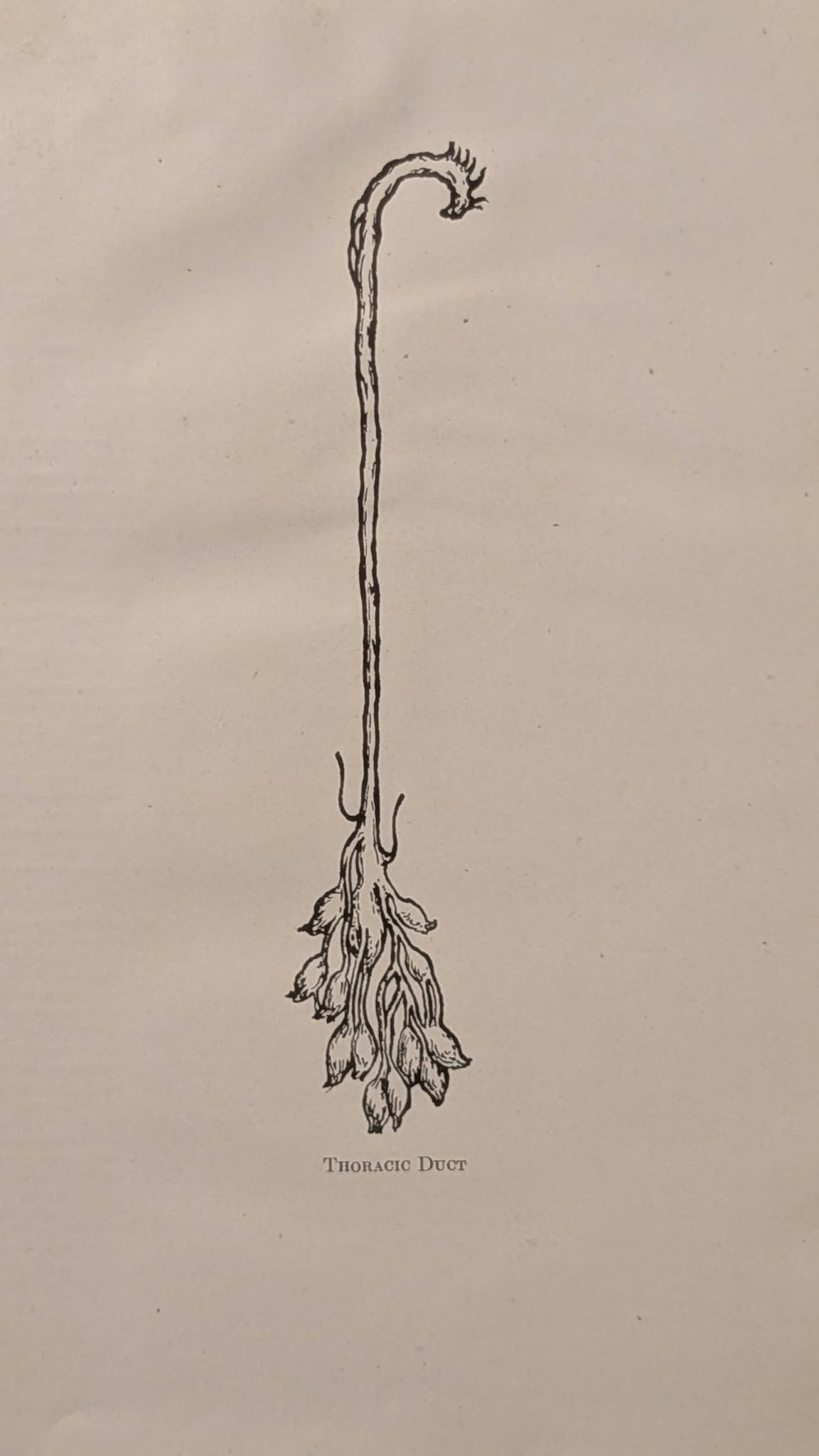
The cisterna chyli receives lymph from the lumbar lymphatic trunks, which drain the lower limbs, pelvis and abdominal wall, together with lymph from the intestinal lymphatic system.
From here, the thoracic duct ascends towards the diaphragm.
However, there is an important anatomical variation that we should understand.
The cisterna chyli is not present as a recognisable structure in everybody.
A 2024 anatomical meta-analysis (1) discussed in the podcast found an identifiable cisterna chyli in approximately 55% of the specimens examined.
In other individuals, the thoracic duct may arise from a convergence of lymphatic channels or a more complex network of interconnected vessels.
This is a useful reminder that anatomical illustrations are representations of common arrangements rather than exact descriptions of every patient.
Anatomical variation is not an error in human anatomy. It is part of human anatomy.
And the lymphatic system demonstrates this particularly well.
The diaphragm has its own specialised lymphatic system
Before following the thoracic duct into the thorax, I want to explore an anatomical relationship that I think deserves much more attention in osteopathy.
We tend to think of the diaphragm primarily as a respiratory muscle.
We study its attachments, innervation, movement and relationship with the thoracic and abdominal cavities.
But the diaphragm also contains an extensive lymphatic network.
On its abdominal surface, immediately beneath the peritoneal lining, are specialised lymphatic structures involved in the absorption of material from the peritoneal cavity.
These include lymphatic lacunae and microscopic openings known as stomata.
What are diaphragmatic lymphatic stomata?
The abdominal surface of the diaphragm is covered by peritoneum. Within particular regions of this mesothelial surface are microscopic openings between cells called stomata.
These openings provide communication between the peritoneal cavity and the lymphatic structures beneath it.
The anatomical arrangement can be understood as a pathway from the peritoneal cavity through the mesothelial stomata into short subperitoneal channels and then into the underlying lymphatic lacunae.
The lacunae are specialised expanded lymphatic spaces that connect with the wider diaphragmatic lymphatic network.
This system is capable of absorbing more than fluid alone. Experimental studies have demonstrated that particles and cells can pass from the peritoneal cavity through the stomata into the underlying lymphatics.
It is a remarkable anatomical arrangement.
And it becomes even more interesting when we consider where these structures are located.
Respiration and diaphragmatic lymphatic function
The diaphragm is constantly moving.
During inspiration, its muscle fibres shorten and the diaphragm descends. Thoracic and abdominal pressure relationships change.
During expiration, the diaphragm relaxes and rises again.
The lymphatic vessels and lacunae embedded within the diaphragm therefore exist in a continuously changing mechanical environment.
Experimental research suggests that respiratory movement and pressure can influence the morphology and patency of diaphragmatic stomata and their associated lymphatic structures.
This gives us a much more detailed anatomical picture than simply saying that the diaphragm pumps lymph.
The diaphragm contains a specialised lymphatic absorption system within moving tissues that experience changing pressures.
It is a dynamic relationship between respiratory mechanics, pressure and lymphatic physiology.
Importantly, not all lymph absorbed through the diaphragm travels directly into the thoracic duct. Diaphragmatic lymphatic drainage involves regional pathways through mediastinal, parasternal and abdominal lymphatic networks.
We are dealing with a complex system rather than a single drainage pipe.
Embryology explains the unusual course of the thoracic duct
One of the most interesting features of the thoracic duct is its course through the body.
It generally begins towards the right side of the midline, ascends through the thorax, crosses towards the left and eventually reaches the root of the neck.
Why does it follow this unusual route?
The answer becomes clearer when we study its embryological development.
During development, the thoracic duct arises from paired longitudinal lymphatic channels with connections between them.
As development progresses, portions of these channels persist while others regress.
The usual adult thoracic duct is formed predominantly from the lower portion of the embryonic right thoracic duct and the upper portion of the embryonic left thoracic duct, connected through an anastomosis.
This helps explain why the adult vessel crosses from right to left.
It also helps explain why anatomical variations occur.
Depending on which embryological channels persist or regress, the adult thoracic duct may be duplicated, partially duplicated or organised into a more complex network. Its crossing point and termination may also differ.
Embryology does much more than tell us where a structure came from.
It helps us understand why adult anatomy looks the way it does.
Following the thoracic duct through the thorax
The thoracic duct usually enters the thorax through the aortic hiatus of the diaphragm.
It then ascends through the posterior mediastinum, where it has several important anatomical relationships.
The vertebral bodies lie posteriorly. The oesophagus is anterior to the duct, while the azygos vein generally lies to its right and the descending thoracic aorta towards its left.
As it ascends, the duct usually crosses towards the left side of the thorax before continuing towards the thoracic inlet.
Consider the environment through which this small vessel travels.
The oesophagus changes shape during swallowing. The aorta pulsates. The rib cage and thorax change dimensions during respiration and the pressure relationships within the thoracic and abdominal cavities continually fluctuate.
Running through this environment is a thin-walled lymphatic vessel only a few millimetres across.
For me, this is where knowing anatomical relationships becomes much more useful than simply memorising the course of the duct.
Where does the thoracic duct terminate?
The classical anatomical description places the termination of the thoracic duct at the left venous angle, also known as Pirogoff's angle.
This is the junction between the left internal jugular and left subclavian veins.
However, the termination is remarkably variable.
The 2024 anatomical meta-analysis discussed in the podcast found that the left venous angle was the most common termination but accounted for approximately 45% of pooled cases.
Other arrangements include termination into the internal jugular, subclavian or brachiocephalic veins.
The terminal duct may also divide into multiple branches.
These variations are particularly important in procedures involving the lower neck, oesophagus and thorax because injury to the thoracic duct can result in lymphatic leakage.
Within the thorax, this may cause a chylothorax, where chyle accumulates within the pleural cavity.
Because chyle contains absorbed dietary fats, proteins, electrolytes and immune cells, a significant persistent leak can have nutritional, immunological and respiratory consequences.
Anatomical variation is therefore much more than an interesting detail in a textbook.
It can have important clinical consequences.
What actually moves lymph through the thoracic duct?
This is where the physiology becomes particularly interesting from an osteopathic perspective.
The traditional explanation of thoracic duct flow emphasises respiration.
As the diaphragm descends during inspiration, abdominal pressure rises while thoracic pressure falls. These changing pressure relationships can contribute to lymph movement towards the venous circulation.
Valves help limit backward flow.
There is physiological support for the influence of respiration on lymphatic flow, but the mechanism is more complex than this simplified model suggests.
A systematic review published in 2026, (2) discussed in the podcast, examined thoracic duct pressure and flow in human and animal studies.
Respiration influenced thoracic duct flow or pressure in most of the human studies and many of the animal studies. However, the findings were not entirely consistent and the magnitude of the effects varied.
The review also highlighted another important mechanism.
The thoracic duct is not simply a passive pipe
The wall of the thoracic duct contains smooth muscle.
This means that the vessel can generate its own contractions rather than relying entirely on external mechanical forces to move lymph.
Segments of lymphatic vessels can actively contract while valves help organise the direction of flow.
Respiration, cardiovascular activity and movement can interact with this intrinsic contractile activity.
There is also evidence that the contractile behaviour of the thoracic duct is influenced by neural and chemical mechanisms.
So rather than imagining lymph being pushed through a passive drainage tube, we should understand the thoracic duct as living vascular tissue participating in an active physiological system.
This is an important distinction when we begin considering the potential influence of osteopathic treatment.
Can osteopathic treatment influence lymphatic flow?
The relationship between osteopathic treatment and lymphatic physiology has been a subject of interest within the profession for many years.
Given the anatomy we have explored, it is reasonable to ask whether changing respiratory mechanics, movement or pressure relationships might influence lymphatic function.
And there is experimental evidence that certain mechanical interventions can influence lymphatic flow.
Animal studies have directly measured thoracic duct flow during osteopathic lymphatic pump techniques and demonstrated significant increases.
In one canine experiment discussed in the podcast, abdominal lymphatic pump treatment increased measured thoracic duct flow from approximately 1.6 to 4.8 millilitres per minute.
Thoracic lymphatic pump treatment increased flow from approximately 1.2 to 3.45 millilitres per minute.
Other experimental studies have demonstrated increased thoracic duct lymph flow and increased movement of leukocytes through lymph during lymphatic pump treatment.
These findings are interesting because they demonstrate that mechanical intervention can influence lymphatic physiology under experimental conditions.
However, we must distinguish those findings from what we can currently establish in human osteopathic practice.
An experimental increase in thoracic duct flow in an animal does not establish that an individualised osteopathic treatment produces the same physiological response in a human patient.
Nor does it establish whether a measured change in lymphatic flow produces a meaningful clinical benefit.
These are questions that require further investigation.
What would better research look like?
Modern lymphatic imaging is creating opportunities to investigate questions that were previously extremely difficult to study.
Techniques such as magnetic resonance lymphangiography allow researchers to visualise central lymphatic anatomy and investigate lymphatic flow in living patients.
This opens up some fascinating possibilities.
Could we measure thoracic duct flow before and after a particular osteopathic intervention?
Could we investigate whether changing respiratory mechanics alters lymphatic movement?
Could we examine how different breathing patterns influence central lymphatic flow?
And if physiological changes occur, do they have meaningful clinical consequences?
These are the sorts of questions I would love to see investigated.
As clinicians, we can observe changes in a patient's symptoms, movement, breathing or function following treatment.
Those observations are useful clinical information.
But a patient's improvement does not necessarily tell us which physiological mechanism produced that improvement.
Clinical observation and experimental research should inform one another rather than compete.
What does the thoracic duct teach us about osteopathic thinking?
After exploring all of this anatomy and physiology, I don't come away thinking that every osteopath needs to learn a specific thoracic duct technique.
Quite the opposite.
I come away thinking about relationships.
The thoracic duct connects anatomical regions that we often learn separately.
It passes from the abdomen through the diaphragm and posterior mediastinum, along the vertebral column and into the root of the neck.
Along the way, it exists in close relationship with the oesophagus, azygos venous system, aorta and other mediastinal structures.
Its function is influenced by a combination of intrinsic contractility, pressure relationships and external mechanical influences.
And the lymph it transports is involved in fluid balance, nutrition and immune function.
This is why I find the thoracic duct so interesting osteopathically.
It reminds us that the body doesn't operate according to the separate chapters in our anatomy textbooks.
We study the musculoskeletal, cardiovascular, digestive and lymphatic systems separately because it helps us learn.
But in the living patient, these systems exist together.
Their anatomical and physiological relationships matter.
The thoracic duct provides a particularly interesting example of that integration.
Final thoughts: Anatomy gives us questions worth asking
The more closely I have studied the thoracic duct, the less it resembles a passive drainage pipe and the more it looks like part of a living, active and remarkably variable system.
Next time you examine the diaphragm, don't just think about it as a respiratory muscle.
Think about what lies above it, what lies below it and what passes through it.
Think about the lymphatic networks embedded within its tissues and the microscopic stomata on its peritoneal surface.
Think about how respiratory movement changes the mechanical environment in which these structures exist.
And think about the questions these relationships raise for osteopathic practice.
Could changing respiratory mechanics influence lymphatic physiology? Could osteopathic treatment influence some of these processes? How might we investigate those possibilities more rigorously?
Some of these questions already have experimental evidence behind them. Others remain hypotheses that require further investigation.
That is exactly why they are worth exploring.
Anatomy gives us relationships. Those relationships give us questions. Our patients give us observations. And research gives us another way of testing and refining what those observations might mean.
That is a process I believe osteopathy should be part of.
Because the more detail we learn, the less the body looks like a collection of separate structures and the more it begins to look like a series of relationships.
By Jamie Archer | The LXO
_____________________________________________________________________________________________________
The Thoracic Duct: The Anatomy We Probably Don't Think About Enough
The LXO Podcast with Jamie Archer
Explore the anatomy and physiology discussed in this article in Jamie's podcast episode, including the research into lymphatic flow and its relevance to osteopathic practice.
_____________________________________________________________________________________________________
References:
1. Anatomical meta-analysis
Plutecki D. et al. (2024). The Anatomy of the Thoracic Duct and Cisterna Chyli: A Meta-Analysis with Surgical Implications Journal of Clinical Medicine, 13(15), 4285.
DOI: 10.3390/jcm13154285
2. Physiological systematic review
Moazzam et al. (2026). The physiology of thoracic duct pressure and flow: A review of the literature - Moazzam - 2026 - Physiological Reports - Wiley Online Library Physiological Reports, 14(2), e70742.
DOI: 10.14814/phy2.70742
Stay connected with news and updates!
Join our mailing list to receive the latest news and updates from our team.
Don't worry, your information will not be shared.
We hate SPAM. We will never sell your information, for any reason.